


Exercise Interventions for Tendinopathy: What the Evidence Now Tells Us

Updated: Sep 4
Tendinopathy is among the most persistent and clinically frustrating conditions in athletic practice. It accounts for a disproportionate share of chronic overuse injury, and its management has undergone significant revision over the past decade as the underlying biology has become better understood. While the principles discussed here apply across tendons throughout the body — from the patellar and gluteal tendons to the extensor tendons of the forearm — the Achilles tendon provides the most instructive model.
The reason is straightforward: the Achilles tendon is responsible for approximately 30% of all overuse sports-related injuries. As Bojsen-Møller and Magnusson (2015) have described it, it is the only tendon in the body that operates close to its failure rate on a daily basis in athletic populations. During uphill running and jumping, it is exposed to loads of up to 12 times bodyweight. The margin between what the healthy Achilles tendon tolerates and what causes it to begin failing is remarkably narrow. Understanding why it fails — and more importantly, how to rehabilitate and protect it — requires a clear picture of tendon biology, muscle architecture, and the central nervous system's role in pain and motor control.
Tendon Biology: Structure, Energy Storage, and the Fascicular Problem
Tendons are composed primarily of Type 1 collagen fibers, which account for 70 to 80% of dry tendon mass. These fibers are organized into fibrils, bundled into fascicles, and held together by an extracellular matrix that permits controlled inter-fascicular sliding. This architecture is precisely calibrated for the tendons' primary athletic function: storing and returning elastic energy during gait.
The tendons distal to the knee and elbow — the Achilles, patellar, and plantar fascia among them — are energy-storing tendons. Their associated muscle bellies are relatively short and fire isometrically just before peak force application during running and jumping, locking down and allowing the tendons to lengthen by as much as 11% and then snap back. This spring-like mechanism contributes approximately 60% of the total work of the muscle-tendon complex during running — a contribution that no shoe technology currently available comes close to replicating. By contrast, so-called positional tendons — the gluteus medius, piriformis, and gluteus maximus tendons — have large muscle bellies with short tendons that stretch only about 2%. Their function is force transmission into the joint, not energy storage. The distinction matters enormously for how each type of tendinopathy is managed.
A clinically important architectural feature of the Achilles tendon is the differential rotation of its three contributing muscles. The medial gastrocnemius rotates approximately 28 degrees before inserting into the tendon; the lateral gastrocnemius rotates 136 degrees; the soleus rotates 129 degrees. Medial gastrocnemius force is therefore transmitted almost directly into the tendon, while lateral gastrocnemius and soleus forces must uncoil through their rotational pathway — a process that itself constitutes a form of elastic energy storage. The medial gastrocnemius, because of its more direct line of pull, is also perpetually active during static stance as the primary anti-fall muscle, making it chronically tighter and more prone to limiting ankle dorsiflexion. Restriction of medial gastrocnemius extensibility has been linked to a five-fold increase in forefoot stress fractures, interdigital neuromas, and hallux valgus.
Perhaps the most clinically consequential structural fact about tendons is this: tendon fascicles have no crosslinks between them, unlike muscle fibers which distribute load laterally through spanning myofibers. When a subset of muscle fibers are consistently recruited — as occurs with injury, fatigue, or poor motor patterns — the corresponding tendon fascicles are repeatedly loaded while adjacent fascicles receive no tensile stimulus at all. Tendon fascicles require mechanical loading to stay healthy; unloaded fascicles begin to degrade. This is the microstructural mechanism underlying focal tendinopathy, and it is the reason rehabilitation strategies must actively recruit the full complement of contributing fibers rather than simply strengthening the most accessible motor units.

Why Heavy-Load Eccentrics Are No Longer the Gold Standard
"Eccentric vibration does not reliably stimulate the cellular repair processes necessary for tendon recovery.
What does stimulate those processes has become substantially clearer through recent research, and it centers on two mechanisms: interfascicular gliding and isometric loading in a lengthened position."
For decades, the Alfredson heavy-load eccentric protocol (1998) — and the related work of Stanish from the 1970s — dominated tendinopathy management. The protocol was widely adopted and produced reasonable results in many patients. However, a landmark follow-up study found that 60% of patients treated with heavy-load eccentrics still reported pain and discomfort five years after treatment (Michaud, 2025). Multiple studies have since confirmed that heavy eccentric loading not only fails to reliably resolve tendinopathy but can in some cases worsen symptoms (Michaud, 2025). Even researchers originally associated with the Alfredson protocol have shifted their clinical practice toward isometric-dominant approaches.
The original rationale for eccentrics rested on a proposed vibration mechanism: the active tissue lengthening during an eccentric contraction was thought to generate a micro-vibration that stimulates tenocyte remodeling. This hypothesis has not held up under scrutiny. Eccentric vibration does not reliably stimulate the cellular repair processes necessary for tendon recovery.
What does stimulate those processes has become substantially clearer through recent research, and it centers on two mechanisms: interfascicular gliding and isometric loading in a lengthened position.
The Science of Tendon Remodeling: Isometrics and Interfascicular Gliding
A 2022 study from Australian researchers examined how fluid moves within tendons during exercise (reviewed in Michaud, 2025). Using imaging technology, they demonstrated that isometric contractions create a hydraulic pumping effect — fluid is pushed out of the tendon under pressure, generating a shear force that directly stimulates tenocytes to initiate remodeling. This was the first mechanistic explanation for why isometric loading produces tendon adaptation, and it shifted the evidence base decisively away from the eccentric paradigm.
Subsequent research established the parameters needed to trigger this remodeling response (Michaud, 2025). Sustained isometric contractions at moderate-to-high intensity combined with adequate time under tension are necessary to produce meaningful reduction in tendon volume — a marker of healthy remodeling — and to activate the downstream cellular machinery. A 40-second isometric contraction, held in a lengthened position, initially loads the healthy tendon fascicles before recruiting the damaged region as the healthy fibers take up their share of the strain and move out of the direct loading path. The pathological section — which under standard eccentric protocols would remain effectively stress-shielded — is thus exposed to therapeutic tensile loading for the first time.
The timing of the remodeling response is also now better understood. Research has identified a 10-minute window during which lysyl oxidase and ERK1/2 — the enzymes responsible for stimulating collagen crosslink formation — are upregulated in response to mechanical loading. Beyond 10 minutes, these enzymes return to baseline. This is why short, 10-minute exercise protocols repeated every 6 to 8 hours are recommended in clinical populations, rather than prolonged single sessions that exhaust the biochemical signaling window after the first few minutes of loading (Michaud, 2025).
Alongside isometric loading, the quality of interfascicular gliding — the relative sliding of tendon fascicles against one another — is emerging as a key determinant of tendon health. A 2021 study demonstrated that exercises producing the greatest interfascicular movement were the most effective for improving tendon remodeling outcomes (reviewed in Michaud, 2025). This means that exercise selection for tendinopathy must prioritize movements that actively drive different fascicles to slide against each other, not merely overall contractile output of the muscle. In practice, this favors bent-knee soleus exercises over straight-leg protocols for mid-portion Achilles tendinopathy, because the bent-knee position places the soleus — the largest contributor to the Achilles complex by muscle mass — in a lengthened configuration that enhances both the range and quality of fascicular gliding through the rehabilitation range of motion.
Full triplanar range of motion through inversion and eversion during rehabilitation is equally essential for recruiting the complete fascicular breadth of the Achilles tendon. Bojsen-Møller and Magnusson's (2015) research demonstrates clearly that exercises performed in a single sagittal plane leave large portions of the tendon unloaded and therefore unstimulated. The rear foot must actively invert and evert through the range of rehabilitation exercises — whether isometric or isotonic — to distribute the remodeling stimulus across the full cross-sectional area of the tendon. This is a likely contributor to the incomplete long-term outcomes of the Alfredson protocol, which is performed exclusively in the sagittal plane.
The Central Nervous System Dimension: Cortical Inhibition and Its Consequences
One of the most underappreciated dimensions of tendinopathy management is its neurological component. When a tendon becomes painful, the central nervous system responds with cortical inhibition — a protective downregulation of motor output to the affected region. Transcranial magnetic stimulation studies have localized this inhibition to specific cortical areas, confirming that it is a genuine neurophysiological phenomenon, not merely a behavioral avoidance response (Michaud, 2025). The neurotransmitter GABA is reduced in the inhibited pathways; descending inhibitory signals suppress motor unit recruitment in the injured limb — and, remarkably, in the contralateral limb as well. Athletes with unilateral Achilles tendinopathy show a 38% reduction in calf endurance on the symptomatic side and a 34% reduction on the asymptomatic side (Michaud, 2025). This bilateral pattern is the most compelling evidence available that the impairment is driven by central inhibition rather than by isolated tissue damage alone.

Rio and colleagues' (2015) research established that sustained isometric contractions are an effective clinical tool for reducing cortical inhibition and managing acute tendon pain. In their studies of patellar tendinopathy, a single 45-second isometric contraction at approximately 70% of maximum voluntary effort produced an immediate reduction in pain from an average of 6.8 to 2.6 on a 10-point scale, with pain relief persisting for up to 45 minutes. The mechanism appears to involve a reduction in the descending inhibitory signal, with a corresponding increase of up to 20% in measured muscle strength. For athletes managing a painful tendon through a competitive season, a series of four to five 45-second isometric contractions before training or competition provides a practical window of pain reduction and motor unit restoration that allows performance without further sensitizing the central nervous system.
A 2024 study introduced another powerful tool for reducing cortical inhibition: metronome-paced exercise (Gordon, Jeanfavre and Leff). When patients performed their rehabilitation exercises synchronized to a metronome, the requirement for precise temporal coordination actively engaged the central nervous system in a way that suppressed inhibition. The external timing cue forced the nervous system to attend to movement rather than pain, and the precision demanded stimulated both strengthening of existing neural connections and potential formation of new ones. In clinical practice, this means setting a metronome to match the athlete's typical movement cadence — for a runner with 180 ground contacts per minute, setting it to 90 beats per minute — and having them perform isometric and subsequently isotonic exercises synchronized to that beat. The progression runs from isometric contractions in the early phase, through isotonic loading in mid-rehabilitation, to plyometric work in later stages, with the metronome present throughout.
"One of the most underappreciated dimensions of tendinopathy management is its neurological component. When a tendon becomes painful, the central nervous system responds with cortical inhibition — a protective downregulation of motor output to the affected region."
Load Calibration: The Critical Role of Ligamentous Laxity
Perhaps the most clinically significant recent advance in tendinopathy management is the individualization of isometric loading intensity. The standard recommendation — 70 to 80% of maximum voluntary contraction — represents a reasonable population average but is physiologically incorrect for a substantial proportion of athletes.
Research using ultrasonography to measure tendon fascicle elongation during loading has established that the optimal mechanical stimulus for tendon remodeling requires approximately 6.2% tendon elongation (Michaud, 2025). The key finding is that achieving this elongation requires dramatically different loads in different individuals. In hypermobile athletes, 6.2% elongation may be reached at only 47% of maximum voluntary effort. In stiff, mesomorphic athletes, reaching the same elongation may require 90% of maximum effort. The implications of getting this wrong are significant: prescribing heavy loads to hypermobile individuals drives excessive fascicle separation — above 9% elongation, injury risk increases 2.3-fold. Prescribing lightweight protocols to structurally stiff athletes produces insufficient strain to stimulate remodeling at all (Michaud, 2025).
A practical clinical screen for ligamentous laxity uses the thumb-radius index: the athlete presses their thumb toward their forearm using the opposite hand. If the thumb approaches within 2 centimeters of the forearm, ligamentous laxity is present and isometric loading should be calibrated to 40–50% of maximum voluntary effort. If the thumb cannot reach within 2 centimeters of the forearm, the athlete is structurally stiffer and can be worked at 70–90% effort to achieve the target fascicle elongation. This single screen takes under 30 seconds and substantially narrows the error range in load prescription.
Synergist Strengthening and Variable Load Transfer
A comprehensive Achilles tendinopathy program extends beyond the gastrocnemius-soleus complex to address the full network of muscles that share the mechanical load of plantar flexion. This concept — variable load transfer — recognizes that the Achilles tendon's load at any given moment reflects not only what it is being asked to produce directly, but also what its synergists are failing to contribute.
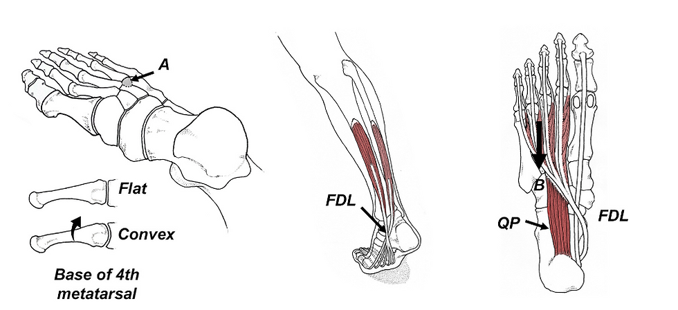
Two muscles deserve particular emphasis. Flexor hallucis longus, attaching from the fibula to the distal phalanx of the great toe, is a powerful plantar flexor with a substantial lever arm to the ankle axis. When it is weak, the Achilles tendon must compensate by absorbing forces that flexor hallucis longus would otherwise handle. MRI studies of athletes with Achilles tendinopathy consistently show hypertrophy of the flexor hallucis longus three months after injury — the body's own evidence of its attempt to redistribute load away from the damaged structure. Directly strengthening the great toe flexors as a component of Achilles rehabilitation is not counterintuitive; it is mechanically rational and clinically supported.
Peroneus longus, wrapping from the fibula around the lateral ankle and inserting at the base of the first metatarsal and medial cuneiform, is the primary stabilizer of the medial forefoot and a powerful plantar flexor. Its Y-shaped attachment provides a compressive locking function at the first tarsometatarsal joint; weakness allows the first ray to become hypermobile, transferring excess stress into the Achilles and increasing the risk of forefoot pathology. Toe-in heel raises activate peroneus longus preferentially, as does any exercise requiring active medial forefoot loading. Both muscles should be specifically targeted in a rehabilitation protocol, not left to incidental recruitment through standard calf exercises.
An Integrated Protocol for Tendinopathy Management
The evidence now supports a rehabilitation approach that integrates four components: prolonged isometric contractions in a lengthened position at an intensity calibrated to the athlete's ligamentous laxity; metronome-paced exercise to reduce cortical inhibition and improve neural drive precision; synergist strengthening targeting peroneus longus and flexor hallucis longus for variable load transfer; and full triplanar range of motion to recruit the complete fascicular architecture of the tendon. Plyometric loading is introduced only after pain has subsided and cortical inhibition has been sufficiently reduced — ensuring that power is expressed through a remodeling tendon rather than one that remains neurally suppressed.
The most important shift in contemporary tendinopathy management is the recognition that the central nervous system is always involved, that pain inhibits performance bilaterally and not only locally, and that tendon remodeling requires both the right mechanical stimulus and the right neural environment. Getting both right simultaneously is the clinical difference between an athlete who recovers fully and one who manages a chronic tendon problem indefinitely.
---
Frequently Asked Questions
Q: Why is the Achilles tendon so much more vulnerable to chronic injury than other tendons?
Two reasons stand out. First, the Achilles operates close to its mechanical failure threshold during normal athletic activity — uphill running and jumping can expose it to loads of up to 12 times bodyweight, leaving almost no margin for error when load management is poor. Second, the medial gastrocnemius is perpetually active during static stance as the primary anti-fall muscle, meaning it never fully recovers between bouts of loading the way a muscle recruited only during movement would. This chronic tightness restricts ankle dorsiflexion and alters load distribution through the tendon, creating the conditions for focal tendinopathy over time.
Q: What is wrong with heavy-load eccentric protocols, and why were they used for so long?
The original rationale was plausible — eccentric contractions generate micro-vibrations that were thought to stimulate tenocyte remodeling. The problem is that this mechanism has not been confirmed experimentally, and five-year follow-up data show that 60% of patients treated with the Alfredson protocol still report pain and functional limitation. Additionally, standard eccentric protocols are performed in a single sagittal plane, which leaves large portions of the tendon's fascicular cross-section unloaded and unstimulated. They also fail to address the cortical inhibition component of tendinopathy, which limits their effectiveness regardless of the mechanical stimulus applied.
Q: How does the thumb-radius test change my treatment approach?
It changes the load prescription substantially. Hypermobile athletes who reach the 6.2% target fascicle elongation at only 47% of maximum effort will not benefit from — and may be harmed by — standard 70–80% isometric protocols. Above 9% fascicle elongation, injury risk increases 2.3-fold. The test takes under 30 seconds and gives you a clinically meaningful indication of where to start. Lax individuals work in the 40–50% effort range; structurally stiffer individuals need 70–90% to reach the same target elongation. Without this screen, a single load prescription applied to a mixed population will be undertreating some athletes and overloading others.
Q: Why does Achilles tendinopathy cause weakness in the unaffected leg?
Because the impairment is mediated in part by the central nervous system, not only by local tissue damage. When the tendon becomes painful, descending inhibitory signals from the brain suppress motor unit recruitment bilaterally — athletes with unilateral Achilles tendinopathy show a 38% reduction in calf endurance on the symptomatic side and a 34% reduction on the asymptomatic side. This bilateral cortical inhibition is the reason that rehabilitation must address the neurological component — specifically through isometric contractions that reduce inhibition and metronome-paced exercise that forces the central nervous system to re-engage — and not only the mechanical loading of the tendon itself.
Q: When is it appropriate to introduce plyometric loading in Achilles tendinopathy rehabilitation?
Plyometric loading should only be introduced after two conditions are met: pain has subsided sufficiently that the central nervous system is no longer in a high-inhibition state, and the isometric and isotonic phases have been completed successfully with appropriate load progression calibrated to the athlete's laxity profile. Introducing plyometrics too early does not accelerate recovery — it risks expressing power through a tendon that is still neurally suppressed and mechanically underrremodeled, which is one of the primary mechanisms of re-injury in this population. The metronome should remain in use through the plyometric phase to maintain the neural engagement that suppresses cortical inhibition during the most demanding loading conditions.
References
Alfredson, H., Pietilä, T., Jonsson, P. and Lorentzon, R. (1998) Heavy-load eccentric calf muscle training for the treatment of chronic Achilles tendinosis. The American Journal of Sports Medicine, 26(3), pp. 360–366.
Bojsen-Møller, J. and Magnusson, S.P. (2015) Heterogeneous loading of the human Achilles tendon in vivo. Exercise and Sport Sciences Reviews, 43(4), pp. 190–197.
Cook, J.L. and Purdam, C.R. (2009) Is tendon pathology a continuum? A pathology model to explain the clinical presentation of load-induced tendinopathy. British Journal of Sports Medicine, 43(6), pp. 409–416.
Gordon, J., Jeanfavre, M. and Leff, D. (2024) ‘Effects of tempo-controlled resistance training on corticospinal tract plasticity in healthy controls: a systematic review’, Healthcare (Basel), 12(3):1325.
Kubo, K., Kanehisa, H. and Fukunaga, T. (2001) Effects of isometric training on the elasticity of human tendon structures in vivo. Journal of Applied Physiology, 91(1), pp. 26–32.
Michaud, T. (2025) Exercise interventions for the management of tendinopathy. Journal of Contemporary Chiropractic.
Rio, E., Kidgell, D., Purdam, C. et al. (2015) Isometric exercise induces analgesia and reduces inhibition in patellar tendinopathy. British Journal of Sports Medicine, 49(19), pp. 1277–1283.

Tom Michaud is a chiropractor with 40 years of clinical practice and the author of the best-selling books "Human Locomotion" and "Injury-Free Running" besides being the creator of numerous diagnostic tools and exercise products such as the ToePro and Twist Disk.


Comments